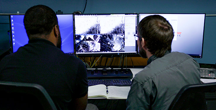
Scanning electron microscopy uses a focused beam of electrons to scan a sample under vacuum. This technique is designed primarily for imaging samples over a range of magnification, typically from ~ <100x to 30,000x.
 The IUP microscopy lab contains a ThermoScientific Prisma E SEM. Designed to be easy to use for a variety of user skill levels, the Prisma E is a versatile research-grade SEM with a tungsten filament, state-of-the-art imaging capabilities, and the ability to host a wide variety of specimens of interest to researchers in myriad disciplines (e.g., anthropology, biology, geology).
The Prisma E includes a Back-Scattered Electron (BSE) Detector, used in compositional imaging (e.g., to distinguish between minerals and glass), and a Secondary Electron (SE) detector, used for topographic imaging, including 3D surfaces. The Prisma E can also be operated under low vacuum or variable pressure conditions (e.g., ESEM mode), which provides the capability to image non-conductive specimens that are too fragile for high vacuum (such as biological specimens), too small to be polished, or cannot be carbon coated (to reduce charging) before observation in high vacuum.
The Prisma E has a large sample chamber (340mm wide), which will hold samples 85mm tall and can view a large area (up to122mm wide) of a sample, due to XY movement with full rotation and up to 90 tilt. The largest field of view is 18mm at the longest working distance. The Prisma E also has high-resolution capabilities (3nm resolution at 30kV under high vacuum) and a magnification range of 6x to 1,000,000x.